Multi-Year Study of 808 Embalmers Across 5 Countries Finds 75.2% Observed White Fibrous Clots in Corpses
The anomalous clots were estimated to be present in 23.4% of all embalmed corpses overall.
This article originally appeared on Focal Points and was republished with permission.
Guest post by Nicolas Hulscher, MPH
A new peer-reviewed study in the International Journal of Innovative Research in Medical Science has officially documented a phenomenon embalmers have been reporting for years: unusual white fibrous structures found in the veins and arteries of deceased individuals.
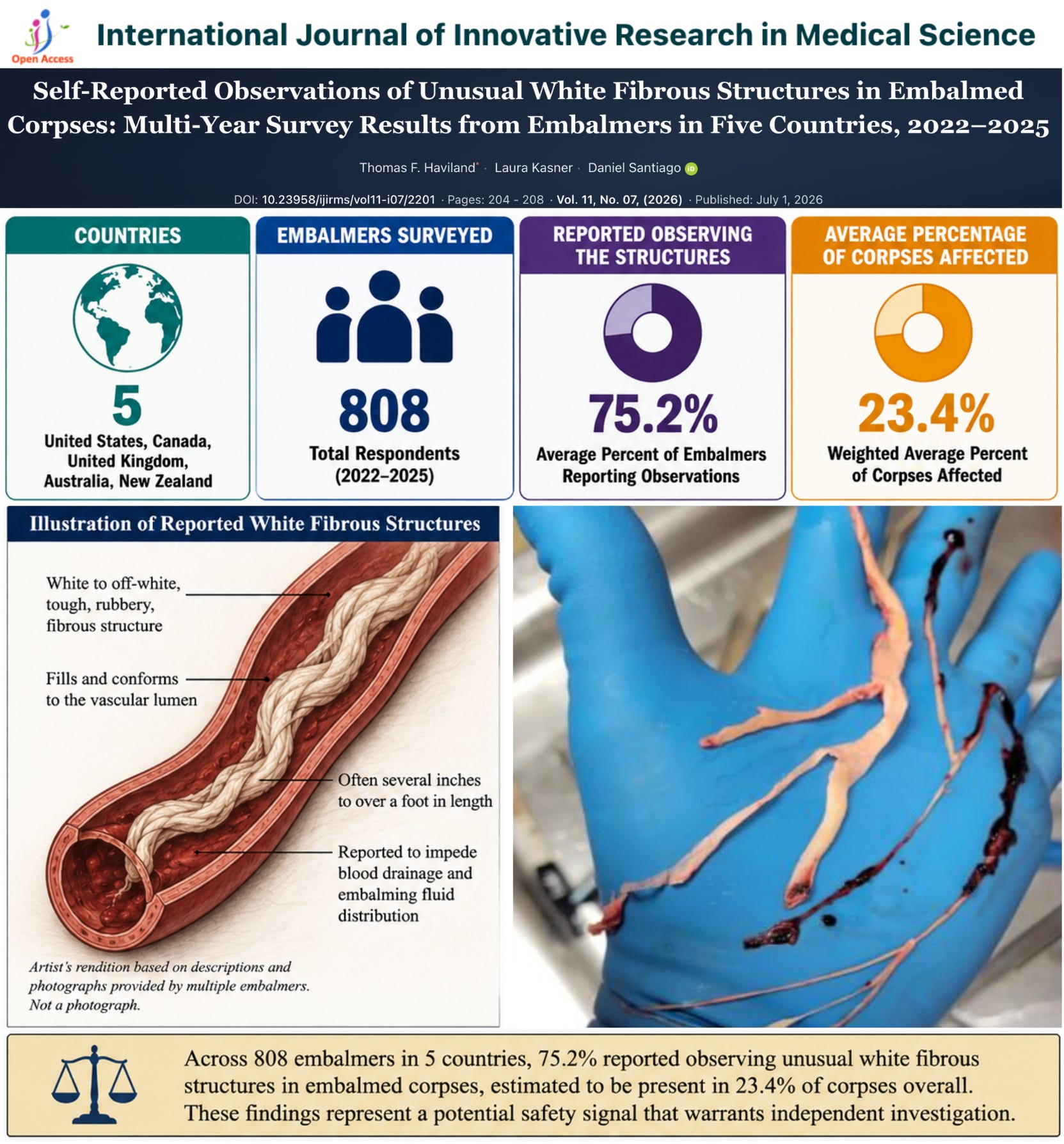
The study was led by Thomas F. Haviland, a retired U.S. Air Force Major, mathematician, and data analyst, along with Laura Kasner and Daniel Santiago, PharmD. Over four consecutive survey years from 2022 to 2025, the researchers collected responses from embalmers in the United States, Canada, United Kingdom, Australia, and New Zealand.
Across the four survey years, 808 embalmers participated.
When the multi-year results are combined, 75.2% of embalmers reported observing white fibrous clots, and the structures were estimated to be present in 23.4% of embalmed corpses overall.


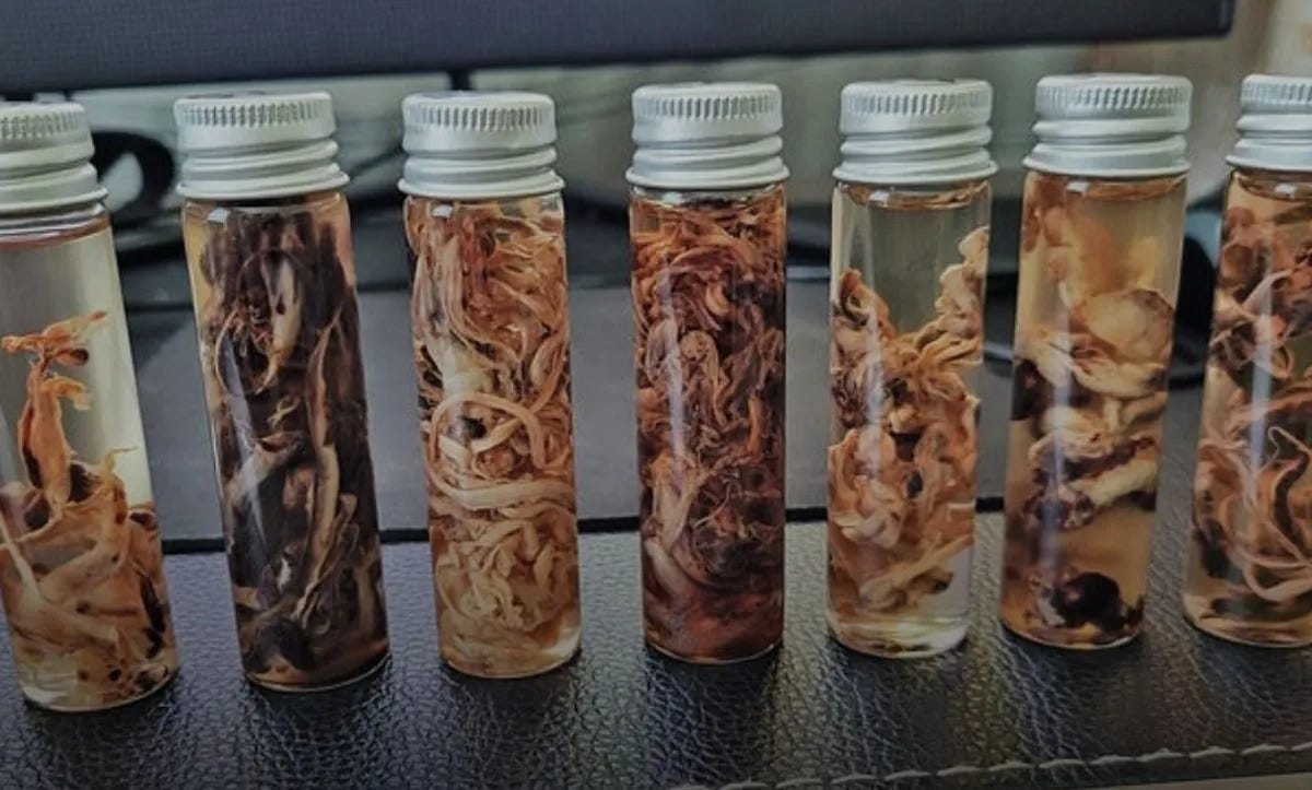
According to respondents, the structures are typically white or off-white, tough, rubbery, and often several inches to over a foot long. Many embalmers reported that they filled blood vessels, obstructed drainage, and interfered with normal embalming fluid distribution. Experienced practitioners described them as distinct from the classic “chicken-fat” and “currant-jelly” clots commonly encountered during embalming.
One of the most notable findings involved timing. In the 2022 survey, embalmers reported a marked increase in first observations beginning in 2020 and accelerating in 2021. This increase coincided with the global rollout of COVID-19 “vaccination” campaigns, during which billions of doses were administered worldwide.
A second paper published the same day went beyond survey data and performed laboratory analysis on representative samples of these fibrous structures. That study was led by Daniel Santiago, PharmD, with Greg Harrison, Miklós Veres, Mark File, and Dennis Planner, and utilized Raman micro-spectroscopy, protein quantification, and amino-acid profiling to characterize the material.
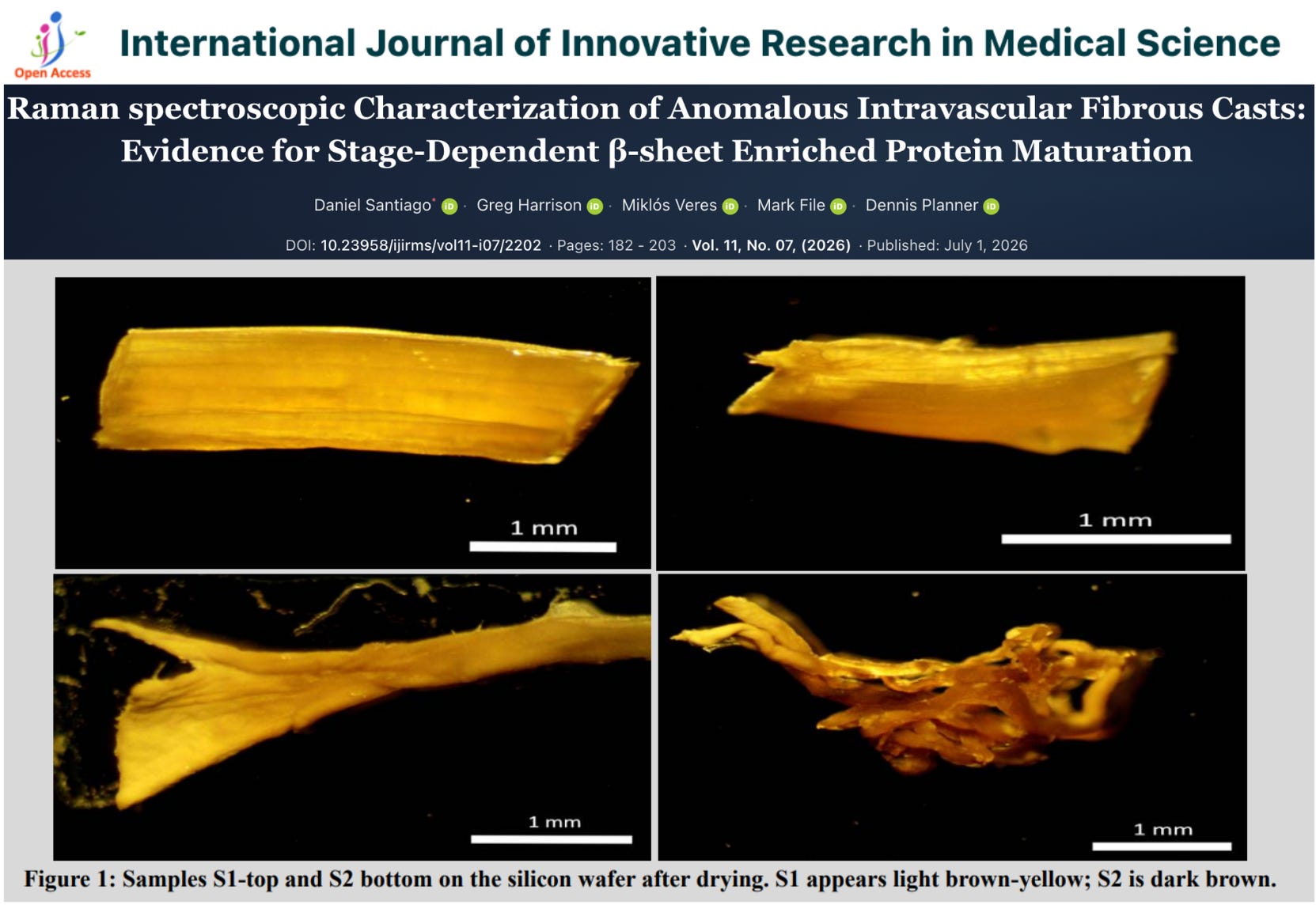
The researchers found strong protein signatures and concluded that the structures appear to represent atypical protein aggregates distinct from conventional postmortem thrombi. Spectroscopic analysis suggested a progression from a native-like α-helical state to a more advanced β-sheet-enriched configuration, consistent with stage-dependent protein aggregation.
In simple terms, the researchers found evidence that these structures are not merely ordinary blood clots. Instead, they appear to consist of abnormal protein aggregates that become increasingly organized and compact over time. This progressive structural maturation may help explain why embalmers frequently describe them as long, rubbery, resilient, and difficult to remove.
While the study found evidence of β-sheet-enriched protein aggregation—one of the hallmarks commonly associated with amyloid formation—the authors emphasized that additional testing is required before the structures can be classified as true amyloid fibrils.
These findings are especially significant when viewed alongside a previously published peer-reviewed study that found amyloid microclots in 100% of vaccinated participants, including every vaccinated “healthy control.”

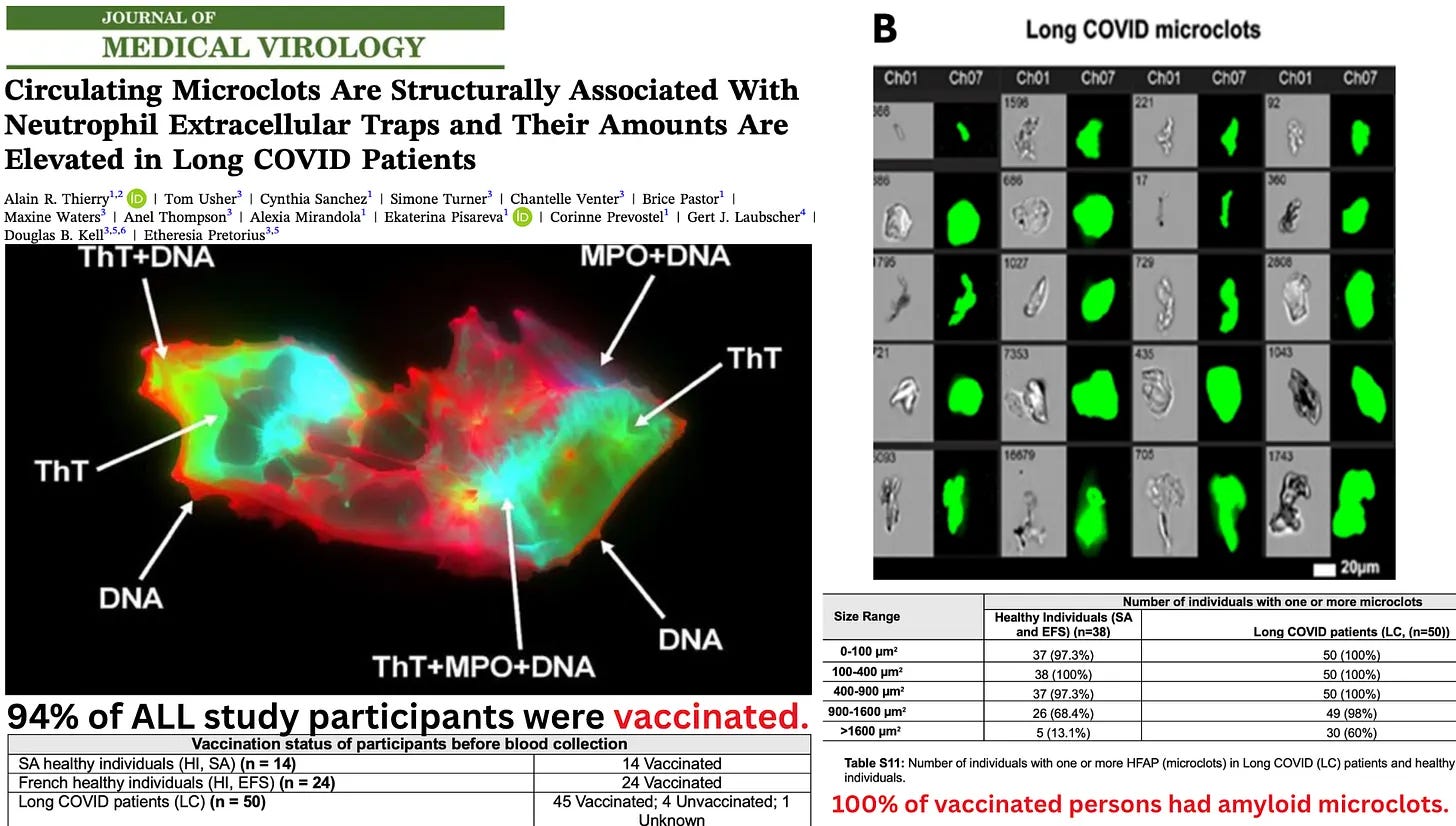
In that study, the pathological microclots were identified using Thioflavin-T, an amyloid-binding dye, and purified spike protein was shown to directly induce the formation of insoluble, fibrinolysis-resistant amyloid aggregates.
Together, these findings raise the possibility that the microscopic amyloidogenic aggregates previously documented in the blood may represent an earlier stage of the same pathological protein-aggregation process that ultimately manifests as the large, rubbery white fibrous casts now being reported by embalmers around the world.
The bottom line is simple: hundreds of embalmers across multiple countries are reporting the same unusual phenomenon, and preliminary laboratory analysis suggests these structures are not ordinary postmortem blood clots.
That must not be ignored any longer.
Epidemiologist and Foundation Administrator, McCullough Foundation
www.mcculloughfnd.org
Please consider following both the McCullough Foundation and my personal account on X (formerly Twitter) for further content.
Copyright 2026 Focal Points